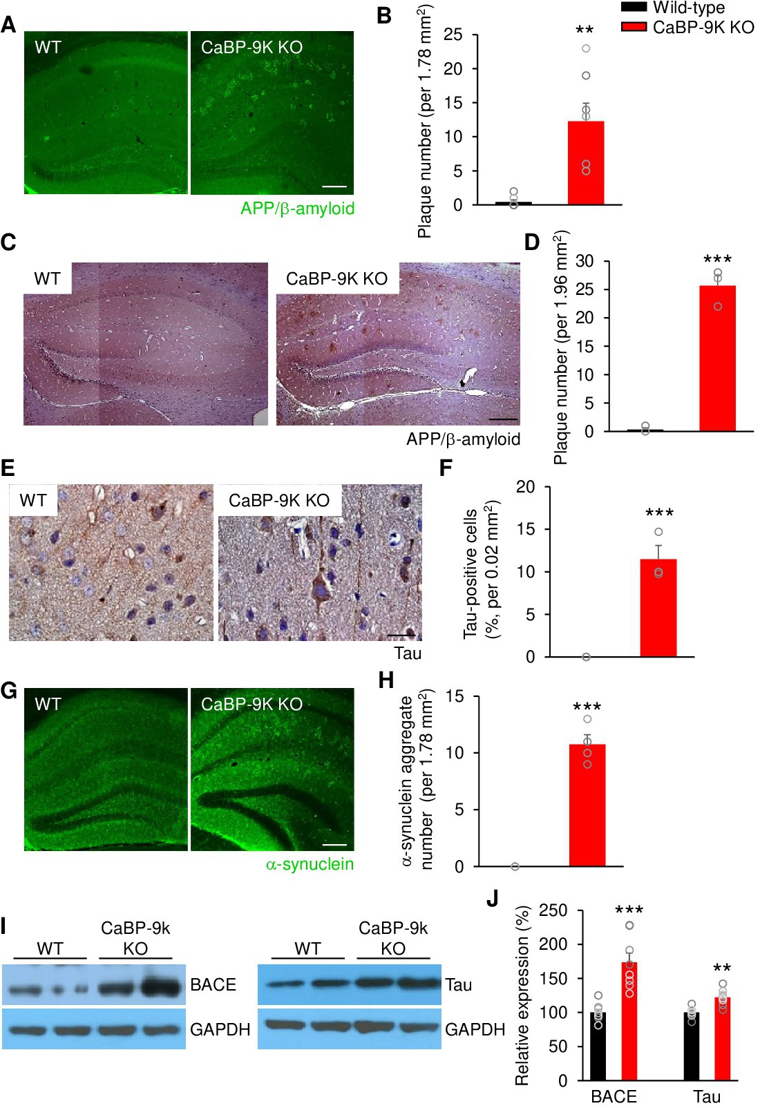
Fig. 1. CaBP-9k knockout causes Alzheimer's disease in mice. (A,C) Hippocampal sections of old wild-type (WT) and CaBP-9k KO mice were stained for APP/β-amyloid by immunofluorescence and immunohistochemistry. Scale bar= 200 µm. (B,D) Quantification of A and C. (Immunofluorescence: n = 7 mice for each group; Immunohistochemistry: n = 3 for mice for each group). (E) Immunohistochemistry for Tau in the hippocampi of old CaBP-9k KO mice. Scale bar= 20 µm. (F) Quantification of E. n = 3 for mice for each group. (G) Immunofluorescence staining of α-synuclein aggregates in the hippocampi of old CaBP-9k KO mice. Scale bar= 200 µm. (H) Quantification of G. n = 4 mice for each group. (I) Western blotting with antibodies to BACE and Tau using brain lysates from old wild-type and CaBP-9k KO mice. (J) Quantification of I. n = 8 for mice for each group. The intensities of the protein bands were normalized to the GAPDH level. Data shown are the means ± SEMs and were analysed by two-tailed unpaired Student's t-tests.